第二届口腔跨学科咬合重建学习班第二届(2014学年)
奥地利Vienna School of interdisciplinary Dentistry -The Slavicek Foundation
福科斯医疗集团
福科斯医疗集团
专家介绍
 Prof. Dr. Markus Greven,MSc,MDS,PhD
奥地利维也纳医科大学牙科学院修复系执行主任
奥地利维也纳医科大学牙科学院修复系“多学科理念修复学”硕士课程执行主任
奥地利维也纳医科大学指定教授
德国法兰克福医科大学牙科学院正畸系正畸硕士教育部Schopf教授合作讲师奥地利维也纳大学多学科牙科学院(VieSID)(主任:R.Slavicek教授):创始成员,顾问组成员,教学组成员
 Dr. Alain Landry
加拿大牙科修复学院成员; 国际正畸协会成员; 美国平衡协会成员; 加拿大牙科协会成员;魁北克牙医协会成员;魁北克外科医生协会会员
2007国际多学科高级牙科学院(IAAID)创始成员
2009VieSID(维也纳多学科牙科学院)教学组成员
VieSID继续教育课程主任
VieSID理论和临床指导教师
 Dr. Anastasia Saltykova
现任Rudolf Slavicek教授的研究研究和科学助理
VieSID牙合学课程临床授课教师
现在奥地利Weber教授人类学研究工作
奥地利维也纳伯恩哈德戈特利布牙科大学门诊主治医生
第二届口腔跨学科咬合重建学习班
当我们把咀嚼器官作为人类机体不可分割的功能部分,咬合的问题就凸显出来了。近年来咬合与其功能的研究被口腔跨学科的研究者所重视,特别在口腔正畸、口腔牙周病、以及种植牙、全口修复重建等。
奥地利Vienna School of interdisciplinary Dentistry -The Slavicek Foundation与中国北京福科斯医疗集团共同举办第二届口腔跨学科咬合重建学习班
理论和操作训练课程分A,B,C,D四个模块,在一年里分别在中国不同地方举办,每个模块5整天,每次都带有操作训练课程,详细内容见下面内容。
(临床病例诊断和分析的高级临床诊断与治疗计划课程在维也纳进行)
授课方式:1.小班上课,提问式互动教学,医生带自己的技师和典型患者共同在课堂上,分析与治疗方案设计。
2.全程中文翻译,
3.带实际病例分析和操作指导.
上课地点 : 北京福科斯医疗集团培训教室
上课时间:Module A: July 9-13, 2014 (5天)
Module B: Oct 24-28, 2014(5天)
Module C: Jan 14-18, 2015 (5天)
Module D: Mar25-29, 2015 (5天)
全部费用:
1.本课程为连续渐进式加深的国际标准课程,四期学习需一次缴纳学费10万¥;
2.凡一次性报名4期基础课程者可提供维也纳2014年7月合学研讨会学费;
3.凡读完4期课程仍然不能很好的应用于临床者可免费读下年课程直至完全掌握;
4.凡因任何原因缺课者不提供补习名额,不授予The Slavicek基金会毕业证书;
报名电话:010-88612600 18611128452
联系人: 郭俊秀 guojunxiu@bjfic.com
基础课程日程安排:
MODULE A : 模块A 牙合学理论基础
Time : 2014.07.09-13 5 Days
Lecturer : Prof. Dr. Markus Greven, MSc, MDS, PhD
Lecture :课程:
- Structures and Functions 结构与功能
The Neuro-Muscular System (NMS) 神经肌肉系统
(including muscle vectors包含肌肉向量)
- Feedback Control System 反馈控制系统
- The Cybernetic system of the masticatory organ (Prof. Slavicek)
- Elements to consider when analysing occlusion : 咬合分析时需考虑的因素
- Posterior support后退支持
- Anterior control 前伸控制
- Lateral control 侧方控制
- Retrusive guidance后退引导
- Vertical dimension of occlusion (V.D.O.)咬合的垂直向高度(V.D.O )
- Sagittal Condylar Inclination (Translation angle of the condyles)
- Transversal Condylar Inclination (Angulation of the Bennett movement)
- Morphologic analysis of the lingual concavity of the upper anterior teeth
- Relative Condylar Inclination相对髁突斜度
- Occlusal plane inclination咬合平面的斜度
- Cusp inclination牙尖斜度
- Curve of Spee Spee曲线
- Curves of Wilson Wilson曲线
- Centric relation正中关系
- Evolution through the ages进化论(随着时间的变化发生的演变)
- Definitions概念
- Reference Position参考位置
- Physiologic 生理的
- Deranged 紊乱的
- Repartition of skeletal Class I, II and III in a given population (Prof. Slavicek’s study)
The “small” clinical functional analysis: “部分”的临床功能的分析
- Clinical exam 临床检查
- Medical anamnesis 大医学病史
- Dental anamnesis 牙科病史
- Occlusal index 咬合指数
- Chief complaint 主诉
- Panoramic X-Ray 全景X射线
- T.M.J. imaging: 颞颌关节成像
- T.M.J. X-Rays taken on panoramic X-Ray machine全景片中的颞颌关节
- What do we really see on T.M.J. x-rays从X片获取T.M.J信息
- Limitations 局限性
- CT-Scans (When, and limitations)CT扫描(何时进行,有何局限性)
- M.R.I. (When, and limitations)磁共振成像(何时进行、有何局限性)
- Muscular palpation 肌肉触诊
- Masticatory system 咀嚼系统
- Neck 颈
- Upper back 上背部
- The 4 minute test: 4分钟测试
- Why, when and how.原因、时间及如何去做
- T.M.J. palpation 颞颌关节触诊
- Lateral pole 侧杆
- Posterior joint space 后部的关节间隙
- Ligament temporo-mandibulare 颞颌韧带
- Mandibular kinematics 下颌运动学
- Dento-skeletal evaluation 牙齿骨骼评估
- Evaluation of facial symmetry 面部对称性评估
- Occlusograms 咬合图
- Evaluation of occlusion with shimstocks 咬合纸测试咬合
- Exact study models: 研究模型分析
- Upper split cast 上颌模型
- Lower Pindex model 下颌模型
- R.P. bite registrations R.P.咬合记录
- Three techniques 3种技术
- Verifying the repeatability of R.P. with an upper split cast model
Bruxcheckers夜磨牙测试
- Discrepancy between Reference Position (R.P.) and inter-cuspal position (I.C.P.)
参考位置(R.R)
- Mechanical Condylar Position Measurement (C.P.M.)
- Resiliency test 髁突弹回力测试
- Myo-functional evaluation功能评定
- Deglutition 吞咽
- Tongue position 舌位置
- Periodontal evaluation 牙周评估
- Soft tissue evaluation 软组织评估
- Primary Brainstem Nerve Analysis 主要的脑干神经分析
- Motor: III, IV, VI, V, VII, XII
- Sensory 知觉的
- V, VII, VIII
- Chronic Pain 慢性痛
- Stress management 精神压力管理
TREATMENT PLAN治疗方案
- Is a Phase I (initial therapy) required? 第一阶段(初步治疗)是否需要?
- Phase II therapy (definitive therapy)第二治疗阶段(确定性地治疗)
- Selective grinding: 选择性调磨
- Indications 适应症
- Contra-indications 禁忌症
- Firstly done on articulator 最初在牙合架上进行
The « small « functional analysis : “部分 ”的功能分析
- Clinical exam (see above), including:临床检查(如下)包括:
- Exact study models:确切的研究模型分析
- Upper split cast 上颌分离模型
- Lower Pindex model 下颌模型
- R.P. bite registrations 参考位置记录
- Three techniques 3种方法
- Verifying the repeatability of R.P. with an upper split cast model
- Mechanical Condylar Position Measurement (C.P.M.)
- Diagnosis 诊断
- Treatment plan 治疗方案

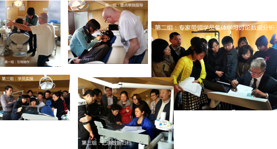
Dr. Alain Landry与学员合作做触诊演示 Prof. Christian现场演示如何上牙合架

MODULE B 模块B: 牙合学数据记录与数据的初步分析
TIME : 2014.10.24-28
Lecture on :
- Large functional analysis, (plus full documentation):强大的功能分析(+全部文档)
- Condylography 髁突轨迹描记图
- Why? 原因
- When?时间
- How?方法
- Electronic condylography 电子髁突轨迹描记图
- How to properly install the hardware 如何正确的安装软件
- How to use the software during the collection of the data (see the I.D.A.L. recommended protocol)
- Condylography 髁突轨迹描记图
- Electronic C.P.M. 电子机械髁突位置测量
- Basic movements 基础运动
- Guided movements 引导运动
- Free movements 自由运动
- Evaluation of the articular capsule 关节囊评估
- Retral stability 横向稳定性
- Transversal Induced Motility 横向动度
- Functional movements 功能运动
- How to register a single reliable R.P. with the aid of the mechanical condylograph
- True Hinge Axis face-bow registration真实的铰链轴面弓记录
- Transferring the reference plane on the skin (to take ceph X-Ray)
- Cephalometric X-Ray头颅侧位片
- Live condylography demonstration (or detailed video presentation to the whole group )
Hands-on, first part : 实操,第一部分
- Clinical hands-on between participants: 4 groups of 5 participants or 5 groups of 4 participants:参加者间进行操作:4组每组5人,或5组每组4人
- Large functional analysis 强大的功能分析
- Electronic condylography 电子的髁突运动轨迹描记
- Cephalometric X-Ray 人头部测量法的X射线
- Large functional analysis 强大的功能分析
Interpretation of condylographic data对髁突运动轨迹描记数据的阐释:
- Quantity, Quality, Characteristics, Symmetry, Reproducibility, Special findings
- Examples of typical condylographic tracings of: 典型髁突运动轨迹描记举例:
- Neuro-muscular system (NMS) 神经肌肉系统(NMS)
- Muscle spasm 肌肉痉挛
- Neuro-muscular disorders 神经肌肉紊乱
- Cranio-Mandibular System (CMS) 颅颌面系统(CMS)
- T.M.J. internal derangements: 颞颌关节内部的错乱
- Luxations 脱臼
- Reducible 可简化的
- Without transversal displacement无横向位移
- With transversal displacement 发生横向位移
- Non-reducible 不可简化的
- Reducible 可简化的
- Luxations 脱臼
- Degenerative Joint Diseases 错乱的关节病
- Arthrosis 关节病
- Arthritis 关节炎
- Neuro-muscular system (NMS) 神经肌肉系统(NMS)
- Morphological analysis of the lingual surface of the upper incisors上切牙舌面形态分析
- Analysis of the occlusal plane on articulator合架上咬合面的分析
- Articulator programming合架设计
- Extra and intra-oral photography 口外和口内照片
- Photography of the casts 模型摄影
- Systematic of case documentation: I.D.AL.documentation protocol病例文档的系统化:I.D.AL.文档协议
Laboratory procedures: 技工室流程
- Mounting the upper cast with True Hinge Axis face-bow 通过真实铰链轴的面弓固定上颌模型
- Mounting the lower cast in R.P. 在参考位置安装下颌模型
- Articulator programming合架操作
- Morphologic analysis of the lingual concavity of the upper anterior teeth上前牙舌面的形态学分析
- Occlusal plane inclination measured on articulator在合架上测量咬合面的斜度
- Interpretation of the condylographic tracings对髁突轨迹描记图的阐释
- Quantity, Quality, Characteristics, Symmetry, Reproducibility, Special findings
- Diagnosis诊断
- Treatment plan治疗方案
- Extra and intra-oral photography 口内外照相
- Photography of the casts 模型摄影
- Case documentation病例记录
- MODULE C 模块C : 牙合学数据的详细分析
TIME : 2015 .1.14-18
LECTURER :
Lecture 课程:
- Interpretation of condylography (more detailed)对髁突运动轨迹描记更详尽的阐释
- Interpretation of tracings:图形阐释
- Quantity, quality, characteristics, symmetry, special findings
- Hyper-mobility 过度移动性
- « loose ligaments »韧带松弛
- Hypo-mobility 动度降低
- Limitations 局限性
- Muscular 肌肉
- Intra-capsular 关节囊内部
- Pain疼痛
- Reducible luxation 可复性盘移位
- Non-reducible luxation 不可复性盘移位
- Acute 急性的
- Chronic 慢性的
- Ankylosis 关节强直
- Degenerative joint diseases 关节紊乱
- Arthrosis, 关节病
- Arthritis 关节炎
- Internal derangements 囊内功能紊乱
- Time curves 时间曲线
- Translation-rotation 转动运动
- Super-imposition of tracings: 过度重叠的曲线
- Protrusion-mediotrusion-opening 前伸-近中移动-开口
- Phonetics-protrusion 发音-前伸
- Deglutition-protrusion 吞咽-前伸
- Bruxism on the protrusion tracing 磨牙症的前伸轨迹图
- Retral bruxism磨牙症的后退运动
- Bruxism in compression 压力下的磨牙症
- Bruxism in distraction 干扰下的磨牙症
- Digitizing the cephalometric points 头影测量数字化定点
- Interpretation of the cephalometric analysis 数字化头影测量分析详释
- Vertical dimension of occlusion咬合的垂直距离
- Compensating mechanisms补偿机制
- Add the protrusive tracing on the ceph.在头影测量分析中加入前伸轨迹
- Interpretation of the cephalometric analysis:头影测量分析数据详释
- Vertical 垂直向
- Occlusal plane angulation and relative condylar inclination
- Protrusion-bruxism 前伸-磨牙症
- Computerized cephalometry数字化头影测量
- Cephalometric analysis 头影测量分析分析
- Dis-occlusion angle and cusp inclination 分离咬合斜度及尖牙斜度
- Angulation of the incisors 切导斜度
- Compensating and decompensating mechanisms:补偿及代谢失调机制
- Dento-alveolar 牙-牙槽骨
- Vertical dimension 垂直距离
- Intra-articular关节内部
- Occlusal plane 咬合平面
Hands-on: 操作
- Condylography, plus full documentation 髁突运动轨迹及全文档记录
- Interpretation of tracings:对轨迹图的阐述
- Computerized cephalometry 数字化头影测量
- Digitizing the cephalometric points 数字化头影测量定点
- Interpretation of the cephalometric analysis对头影测量分析的阐释
- Vertical dimension of occlusion咬合的垂直距离
- Dento-alveolar analysis牙齿-牙槽骨分析
- Occlusal plane 咬合平面
- Functional evaluation功能评估
- Compensating/decompensating mechanisms补偿及代 谢失调机制
Module D 模块 D: 初步的病例计划
TIME;2015.3.25-29
LECTURER :
Lecture 课程:
- Revision of the first modules 对第一模块的回顾
- Including condylographic interpretation 包括对髁突运动轨迹描记的阐释
Phase I therapy for neuro-muscular disorders:对神经肌肉失调的第一阶段的治疗
- Neuro-muscular disorders 神经肌肉失调
- Clinical signs 临床症状
- Options for therapy 治疗的选择
- Type of splint therapy: 夹板治疗的类型
- Emergency splint 急诊颌垫
- R.P. splint 颌垫的参考位置
- Biofeedback splint 颌垫的生物学反馈
- Neuro-muscular, with or without decompression 神经肌肉是否带减压功能
- Verticalization splint 垂直向夹板
- Frequence of controls and adjustments 控制及调整的频率
Phase I therapy for reducible joint luxations:可减少的关节盘移位的第一阶段的治疗:
- Therapeutic postions: 治疗的位置
- Type of splint therapy: 颌垫治疗的类型
- Emergency splint 急诊颌垫
- Arbitrary therapeutic position治疗位置
- Without condylography 无髁突运动轨迹描记
- Disadvantages 缺点
- With condylography 带无髁突运动轨迹描记
- Advantages 优点
- Verticalization splint 垂直向夹板
- Frequence of controls and adjustments 控制及调整的频率
Phase I therapy for non-reducible joint luxations 对不可复的关节移位第一阶段的治疗
- Non-reducible joint luxations 不可减少的关节脱臼
- Acute 急性的
- Clinical signs 临床症状
- Treatment options 治疗选择
- Chronic 慢性的
- Clinical signs临床症状
- Acute 急性的
- Type of splint therapy: 夹板治疗的类型
- Emergency splint 紧急的夹板
- Arbitrary therapeutic position 任意的治疗位置
- With condylography 带有无髁突运动轨迹描记
- Arbitrary therapeutic position 任意的治疗位置
- Decompression splint 减压的夹板
- Verticalization splint 垂直的夹板
- Frequence of controls and adjustments 控制及调整的频率
- Emergency splint 紧急的夹板
Phase I therapy for degenerative joint diseases关节退行性变的第一阶段治疗
- Degenerative joint diseases 关节退行性变
- Arthrosis 关节病
- Arthritis 关节炎
- Treatment options 治疗方案的选择
- Type of splint therapy: 牙合垫治疗的类型
- Emergency splint 急诊牙合垫
- Arbitrary therapeutic position 任意的治疗位置
- With condylography 经髁突运动轨迹描记分析
- Arbitrary therapeutic position 任意的治疗位置
- Decompression splint减压牙合垫
- Verticalization splint 增高牙合垫
- Frequence of controls and adjustments 控制及调整的频率
- Emergency splint 急诊牙合垫
Hands-on: 操作
- Condylography 髁突运动轨迹描记
- Plus full documentation 文档输入
- Diagnosis 诊断
- Phase I therapy (treatment plan, Phase I)第一阶段的治疗(治疗方案、第一阶段)
